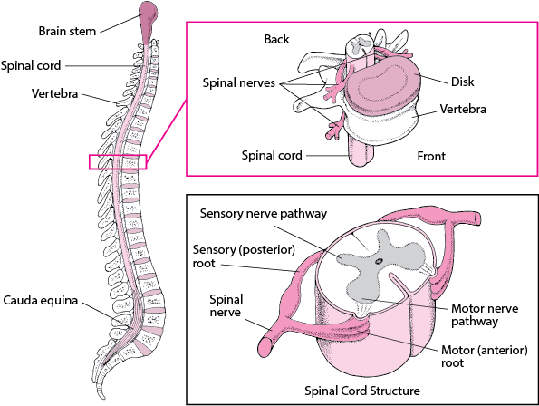
Important structures of the spine

There are a number of important structures in the spine, knowing these structures will help you understand the function of the spine and its related problems. These structures include: intervertebral discs, intervertebral joints (or facet joints), neural foramina, spinal cord, nerve roots, and muscles adjacent to the spine (paraspinal muscles). In the following, we will review each of these sections briefly.
Intervertebral discs (Discs)
Each disc has a strong and hard outer ring made of fiber called Annulus, in which a soft and jelly core called Nucleus Pulposus is placed. The annulus is actually a strong ligament that protects the core of the disc. The nucleus is made up of a tissue that is very moist due to its high volume of water and acts like a shock absorber in the disc structure (something like a mattress filled with water).
Facet joints
Contrary to what many people think, the spine has real joints, similar to knee or elbow joints. In the spine, these joints are called facet joints. Facet joints connect the vertebrae and provide the necessary flexibility for their movement on each other. Between each pair of vertebrae, there are two facet joints. An articular appendage is placed on both sides of each vertebra, and these two appendages are expanded in such a way that they overlap with two articular appendages of the adjacent vertebra to form a facet joint.
Facet joints are movable joints (Synovial Joints). The ends of the bones that reach the movable joints are covered by articular cartilage, which is a soft, spongy substance that allows the bones to slide over each other with minimal friction. Inside these joints, there is a fluid called synovial fluid that keeps the joint surfaces smooth. This liquid is placed inside a waterproof bag made of soft tissue called "joint capsule" and is covered by ligaments that completely surround the joint.
Spinal Cord
The spinal cord is a column of millions of nerve fibers that carry messages from the brain to other parts of the body. The spinal cord extends from the brain to the area between the first and second lumbar vertebrae. The spinal cord is located in a hollow canal called "spinal canal". This channel is created by placing the beads on top of each other. The spinal canal completely covers and protects the spinal cord and its nerve roots.
As mentioned, the spinal cord extends to the second lumbar vertebra. Below this level, the spinal canal contains a group of nerve fibers called the Cuada Equina, which innervate the pelvis and other lower limbs.
The spinal cord is covered by a protective membrane called "Dura Mater" that covers the spinal cord and spinal nerves like a waterproof bag. Inside this bag, the spinal cord is surrounded by the spinal fluid (CSF - CerebroSpinal Fluid). Cerebrospinal fluid protects the spinal cord

Neural Foraminae
The spinal cord branches into 31 pairs of nerve roots, which exit the spine through small openings on either side of the vertebrae (neural foramina). Each pair of nerve roots go in two opposite directions when exiting these openings; One exits from the left orifice and the other exits from the right orifice.
Nerve Roots
As mentioned, nerve fibers branching from the spinal cord form nerve pairs that exit through small intervertebral foramina. The nerves in each area of the spinal cord are connected to specific parts of the body; So that the main reason for paralysis of any part of the body is the injury of the spinal nerves related to the same part.
The nerves of the cervical vertebrae, the upper parts including the chest and hands, the nerves of the thoracic vertebrae, chest and abdomen, and the nerves of the lumbar vertebrae also innervate the legs, pelvis, intestine and bladder. These nerves coordinate all parts of your body, and allow you to control your muscles
Nerve roots are the platform for transmitting nerve messages from the brain to other body organs and vice versa; In other words, if your body is injured, the pain message goes to the brain through these nerves. If these nerves themselves are damaged, you may experience pain, tingling or numbness in the areas of the body that are related to that nerve. Without these nerve messages, your body will not be able to work and will not have a useful function.
Paraspinal Muscles
The muscles adjacent to the spine are called "paraspinal" muscles. In addition to supporting the spine, these muscles are also a stimulus for the movements of this part. Joints help flexibility and muscles help mobility. There are many small muscles in your back and lower back, each of which controls part of the movement of the vertebrae and other parts of the skeletal system